क्या एचएलए-बी 27 परीक्षण जो पहले पॉज़िटिव था वो बाद में नेगेटिव हो सकता है?

क्या ऐसा आपके साथ कभी हुआ है?

आपका  एचएलए-बी 27 परीक्षण जो पहले पॉज़िटिव था वो  बादमें नेगेटिव  हो गया ? क्या आपने ऐसे विज्ञापन देखे है जिसमें कहा गया हो की कोई इलाज से आपका HLA B २७ नेगेटिव कर दिया जाएगा?

तकनीकी रूपसे, यह कभी नहीं हो सकता है । HLA-b27 एक जीन है और रक्तसमूह की तरह यह कभी नहीं बदल सकता है।उस मामलेमें, क्या गलत हो सकता है? 

आइए अब समझते हैं कि एचएलए-बी 27 जीन का परीक्षण कैसे किया जाता है।

HLA B २७ टेस्ट तीन अलग पद्धति से की जाती है—

Microlymphocytotoxicity (MLCT) assay 

Flowcytometry (FC) 

DNA based typing using a Polymerase chain reaction based assay (PCR)

इन विधियों के बीच, पीसीआर आधारित विधि सबसे सटीक/ अच्छी है।

लैब्ज़ अलग अलग पद्धति से HLA B २७ टेस्ट करते है और इसके चलते कभी कभी HLA बी २७ के रिपोर्ट में फ़रक आ सकता है। 

कुछ मरीज़ जो फ्लोसाइटोमेट्री पद्धति से HLA बी २७ नेगेटिव आते हैं;  उनकी टेस्ट पीसीआर पद्धति से पॉज़िटिव आ सकती हैं।

फ़्लोसाइटोमेट्री से कभी कभी रिपोर्ट  अनिश्चित ’(अनिर्णायक) आ सकती है, जिसे पीसीआर आधारित पद्धति से पुष्टि करने की आवश्यकता होती है। तो अगली बार जब आप HLA B २७ टेस्ट करे तब PCR पद्धति से ही करे। लैब से PCR पद्धति की माँग करे।

अगर आपने विज्ञापन देखे है जिनमे इलाज से HLA B २७ नेगेटिव करने की बात कही गयी हो तो उनपर विश्वास नहीं करे । यह मुमकिन नहीं है।

Image courtesy: Free Vectors via http://www.vecteezy.com

Arthritis & COVID-19: Are arthritis sufferers at an increased risk, should medicines be stopped, what specific precautions should be taken?

We are all in difficult time. The numbers affected by COVID-19 are increasing exponentially every other day & we are hearing scary stories from across the globe.

If you suffer from arthritis; I am sure you would have many queries in your mind. Let us try & sort them out today.

1) Arthritis medications work by suppressing the immunity. Does that mean arthritis warriors on prednisolone/ DMARDs/ biologics are at an increased risk?

2) Since medications increase the risk; should the medicines be stopped to mitigate the risk?

3) What would happen if the medicines are stopped? What if the arthritis flares up?

4) Can we calculate the risk based on the medicines & precautions to be taken based on the risk profile?

We have a few videos to answer these questions:


Many of you may have experienced a shortage of hydroxychloroquine (HCQS, Plaquenil) as well. Do you know what you can do if you are not able to procure it?


All in all, there is no reason to panic.

  1. Understand your risk status as per the flow chart in the video
  2. Understand precaution (Standard social distancing, strict social distancing, shielding) & follow them. See what is appropriate for you based on your risk status.
  3. Speak to your rheumatologist. Do not stop any medication on your own.


Image source:

Is there is a shortage of tab. hydroxychloroquine (HCQS, Plaquenil). Why? What can you do if you are not able to procure it?

hcqs

If you are suffering from rheumatoid arthritis/ lupus/ other connective tissue diseases; you must be taking a medicine called hydroxychloroquine (HCQS, Zyq, HQ Tor, OXCQ, Plaquenil). Many of you must not be getting it in the pharmacy stores in recent times.

Why is there a shortage of hydroxychloroquine?

Hydroxychloroquine has been approved in treatment of coronavirus (COVID-19) in many countries across the globe. In India, ICMR has approved it as well. Central & state government in India has procured a substantial stock for the government hospitals. There have been instances of general public buying the medicine as well. All this put together has meant shortage & unavailability in local pharmacies.

Is hydroxychloroquine banned?

No. This rumor has been doing the rounds due to the non availability of the medicine. However, be rest assured, it has not been banned.

What can a patient on hydroxychloroquine do?

Speak to your rheumatolgist. He may be able to help you get the medicine with the help of the pharmaceutical company.

In case you are not able to procure it —

  1. Hydroxychloroquine is a long acting medicine & remains in the tissues for a long time. It is detected in urine even 3 months after stopping it. If you miss it for 7-10 days, there should not be a major issue in a majority.
  2. You may take tab. chloroquine phosphate 250 mg instead of hydroxychloroquine 200 mg. Do speak to your rheumatologist about this before changing over.
  3. Do speak to your rheumatologist & see if he wants to increase the dose of other medicines to make up for hydroxychloroquine.

Pharmaceutical companies have increased the production of hydroxychloroquine & the availability issue should be sorted out soon.

Ref: https://www.accessdata.fda.gov/drugsatfda_docs/label/2017/009768s037s045s047lbl.pdf

Deep breathing exercises for Ankylosing Spondylitis

During normal breathing, ribs move up & down to expand the chest cavity. This movement occurs where they join the vertebrae (costovertebral joint). In ankylosing spondylitis, these joint get inflamed & fused. Once fused, the rib movement gets restricted. This leads to ‘strapped-in’ feeling & breathlessness while doing day today activities.

Just as back exercises keep your spine flexible, so also can deep breathing exercises keep the chest wall elastic & preserve one’s ability to take a deep breath.

Here are a few ‘deep breathing exercises’ to help you keep the chest wall elastic & prevent fusion of the costovertebral joints.


  • Rib cage exercises- standing arm exercise, exercise with the ribcage band, lying down & arm stretch exercises ( first three exercises in the video) help exercise the ribcage muscles & prevent fusion at the costovertebral joints.
  • The next two exercises help exercise the diaphragm. During inspiration, the diaphragm moves downwards & expands the chest cavity. These exercises will help you with a more effective breathing particularly if you already have restriction in rib cage movements.
  • The last exercise will help you understand the ‘rib cage breathing’ & ‘diaphragmatic breathing’ & use both more effectively.

Perform these exercises every day; 10 times at a time, at least twice a day.


One can also use an incentive spirometer to exercise the respiratory muscle.

It is a medical device used to improve the function of the lungs & is generally used after a thoracic surgery. However we can also use it to improve the function of the ribcage muscles.

Here is a video to help you understand how to use it–


Take 15- 20 breaths with the spirometer at least twice a day.

Don’t forget to continue your back exercises. They surely complement these exercises & help you preserve your breathing despite ankylosing spondylitis.

Why is one screened for tuberculosis prior to starting TNF blockers?

Tuberculosis (TB) is an infection caused by bacteria. It most commonly affects the lungs. When a patient of pulmonary TB coughs, the bacteria are discharged in the air. If someone inhales these bacteria, they then find a way into the lungs. Once in the lungs- the bacteria face the following-

  • The bacteria are killed by body’s immune system. They are destroyed & cannot harm the body.
  • They lodge in the body, overcome the immune system & cause an active infection- tuberculosis.
  • The immune system does not kill them but instead jails them (literally; it forms a granuloma—a jail wherein the bacteria remain in dormant- inactive form). So the bacteria are in the body, but are jailed in dormant state & do not cause harm. Anything that frees them from the jail can make them active & lead to an infection.

The bacteria remain in the granuloma jail for years in a dormant phase. They are inactive & do not cause an active infection while in this phase. This is called ‘Latent TB’. The person does not have an active infection & does not spread it to anyone.

TNF alpha is a cytokine (chemical in the body) that is required for maintaining the integrity of the granuloma & also for other defence mechanisms that keep these bacteria dormant. TNF alpha blockers (Remicade, Infimab, Inflectra, Enbrel, Etacept, Intacept, Benepali, Humira, Exemptia, Cimzia, Simponi) are used to treat autoimmune ailments like rheumatoid arthritis, ankylosing spondylitis.

If we start TNF blockers in a person who has latent TB, the granulomas would open up, setting the dormant TB bacteria free & thus leading to active TB infection. This is called for reactivation of TB. This is why, one is always tested for active/ latent TB before TNF blockers are thought of. Active pulmonary TB can be diagnosed by examination & X-ray of the chest along with sputum test. TNF blockers are not given if one has active TB.

Tests for Latent Tb include Mantoux test & IGRA (Interferon gamma release assay commonly known as the TB gold test). A positive test (either) would suggest presence of latent TB.

In case of latent TB, one needs to be treated before TNF blockers can be given. The treatment recommendations vary from country to country. TNF blockers can be given after 1-2 months of this treatment.

Thus the chance of reactivation of TB with TNF blockers is real but can be minimised by proper testing & treatment for latent TB.

Ankylosing spondylitis update from American College of Rheumatology conference 2016


American College of Rheumatology conference is an annual event where experts, Rheumatologists & researchers meet & insights & advances in rheumatological ailments are discussed.

The important updates for AS warriors from the conference is the further research into newer treatment options.

  1. Secukinumab (Cosentyx): has already been approved by US FDA. Studies looking at long term effect (>1 year) were discussed & show good efficacy of the drug in AS.
  2. Tocilizumab (Xeljanz) : A new ray of hope for AS. It has already been approved for rheumatoid Arthritis. It was studied in 207 patients of AS & has shown promising results. However this is quite early. One would require many more studies & the FDA regulatory approval procedure for it to be used in AS. The results definitely look promising & is a ray of hope for AS.
  3. Ustekinumab- another new biologic being studied for AS. Early study shows promising results & is a drug to watch out for.

All in all, a lot of action in the AS treatment arena. With a lot of focus targeted at AS, we should expect more options to treat AS in near future. 

Can a positive HLA-B27 test turn negative later & vice versa?

hla b27

Has this happened to you? A negative HLA-B27 test turning positive later or vice versa?

Technically speaking, this can never happen. HLA-b27 is a genetic marker & just like a blood group it can never change. In that case, what can go wrong?

Let us now understand how the HLA-B27 gene is tested.

It is processed using three different methods

  • Microlymphocytotoxicity (MLCT)
  • Flowcytometry (FC)
  • DNA based typing using a Polymerase chain reaction based assays (PCR)

The methods are arranged in the order of their accuracy with PCR based method being the most accurate. A few patients that test negative with Flowcytometry method do turn out to be positive with the PCR method.

Flowcytometry can also give an ‘indeterminate’ (inconclusive) report that needs to be rechecked with a PCR based method. This is how the discrepancy in subsequent tests sets in.

So the next time to get your HLAB27 checked; do have a look at the method as well.


Image courtesy: Free Vectors via <a href=”http://www.vecteezy.com”>vecteezy.com</a&gt;

Why is breathing difficult & at times, painful in Ankylosing spondylitis?

The mechanical apparatus for breathing consists of the ribs, intercostals muscles, costochondral junctions & the diaphragm. Diaphragm is a muscle located at the base of the chest wall cavity. The ribs move at the joint formed by them with the vertebrae (costovetebral joint). The intercostals muscles are attached to the ribs & are responsible for the movements of the ribs during respiration.

costovetebral joint

During inspiration, the diaphragm moves downwards & expands the chest cavity. At the same time, the ribs move up & out to expand the thoracic cavity. This movement is akin to a bucket handle (moving up). The movement of the ribs is brought about by the contraction of the intercostal muscles.


In Ankylosing spondylitis, there is inflammation of the costovertebral joint. Later, as AS progresses, the joint may also get fused. This inflammation/ fusion then restricts the movement at the joint & consequently the chest expansion. Physically, this manifests as restricted chest expansion.

Ankylosing spondylitis also leads to inflammation at insertion of intercostal muscles to the ribs (enthesitis). This leads to pain in the rib cage area & also further difficulty in taking a deep breath.

Sneezing involves a rapid & high intensity contraction of the intercostal muscles. It is very painful due to the enthesitis & is almost felt like a ‘catch’

Tight control of AS with NSAIDs/ DMARDs/ biologics/ diet & deep breathing exercises are helpful in reducing this chest pain.


What causes foot pain in Ankylosing spondylitis?

Foot pain is a common component of Ankylosing Spondylitis (AS) & other seronegative spondyloarthropathies (SpA- psoriatic arthritis, reactive arthritis). In AS/ SpA, the inflammation is predominantly at two locations- within the joints- synovitis & at the attachment of ligaments/ tendons to a bone (enthesitis)

Let us have a look at the various causes of foot pain—

Heel pain– also known as plantar fasciitis. The foot has a bone called calcaneum located at the heel region. A muscular structure ‘plantar fascia’ is attached at the lower end of this bone.

plantar fascia

There is inflammation at the attachment of this fascia to the bone (enthesitis) & causes pain that is generally maximal in the morning & after periods of rest.

plantar fascitis NSAIDs & at times local steroid injection is used to take care of the fasciitis. Persistent inflammation does indicate ongoing inflammation & would require better control of AS/ SpA.

Use of soft footwear & heel cushion/ cup in footwear helps reduce the pain. A simple exercise involving rolling of the foot on top of a bottle in the morning before walking does a lot good in many.

silicon heel cup

Pain at the backside of the foot– this is commonly due to Achilles enthesitis.

Achilles tendon is the thick band like structure at the backside of the foot. This is attached to the calcaneum bone below.

6Inflammation of the attachment (enthesitis) causes pain & localised swelling.

Copy of 2

Achilles enthesitis is a telltale sign of active AS/ SpA & mandates better control with DMARDs/ biologics.

Toe pain—At times, the entire toe may be swollen & painful. This is known as dactylitis & is a feature of AS.

3

This is suggestive of ongoing active inflammation.

Pain & swelling at the base of the toe can be due to inflammation of the joint & DMARDs/ steroid injection in the joint/ biologics are used to control it.

Mid foot pain– is generally due to inflammation of the underlying joints especially if associated with swelling. This would require better control of AS.

Ankle & midfoot pain can also be due to a flat foot.

5

This would require foot analysis & an insole to correct the flat foot.

foot analysis

All in all, understand your foot pain & discuss with your rheumatologist.

Understanding the reason can help you take better decisions to take control of your AS.

oot pain in AS

Do have a look at a video to understand this better–

How does ankylosing spondylitis cause osteoarthritis of the hip? — the Pacman game

I’m sure all of you know that Ankylosing spondylitis (AS) is an autoimmune disease, which means that the body’s immune system considers its own joints as foreign & attacks them. We have not been able to pinpoint the exact reason for this.
If this immune attack is not controlled in time, it ends up destroying the joints; especially the hips. This is known as osteoarthritis of the hips. Ankylosing spondylitis is a unique disease as it causes destruction of the bones in the hip & at the same time it leads to excessive bone formation in the spine (syndesmophytes). So what starts as backache can well end up as deformities & disability.

But then, how does AS cause destruction of the hips?
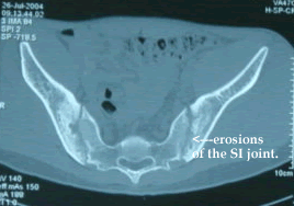
Hip joint is lined by a thin membrane called the synovial membrane from the inside. The immune attack is targeted towards the synovial membrane. The synovial membrane swells up due to the attack. This inflamed synovium is like the pacman. Remember the pacman game?… pacman eats away all the dots. In a similar fashion, the pannus eats the cartilage & the bones around it. When it eats the bones, the same is known as erosion. Apart from the hips, these erosions are also seen in the sacroiliac joints. Destruction of the cartilage & the bone ultimately leads to osteoarthritis.


Rheumatoid_Arthritis_Knee_Joint


pacman

The Pacman game.. Pacman keeps eating the dots…just like the inflamed synovium keeps eating the cartilage & the bones. Note how the AS Pacman has destroyed the hip bones (above) & the sacroiliac joint (SI joint) (below).


Ankylosing_Spondylitis_CT_Scan

Can the hip osteoarthritis be prevented?
Yes, very much so. Seeing a Rheumatologist at the earliest & starting definitive treatment at the earliest can definitely tame this pacman & prevent osteoarthritis.